Key facts
- Stanford University researchers replaced a portion of mouse cortex with human brain organoid cells.
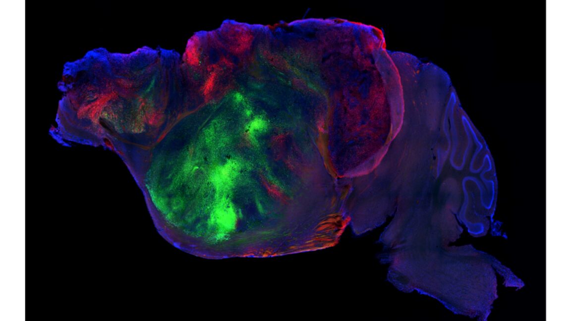
- Human cells integrated into the mouse brain, forming 92% of the cells in the cortex region.
- The human-replaced cortex showed some local organization but lacked distinct layers.
- Mice with humanized cortices performed better than chance on memory tests but not as well as normal mice.
- The humanized cortex mice showed intermediate fine motor coordination compared to normal and cortex-free mice.
Researchers at Stanford University have developed a method to study human brain organoids in a more integrated context by replacing a portion of a mouse's cortex with human brain cells. The team genetically induced the deletion of most cortical cells in developing mice, then implanted human cortex organoids into the resulting void. This allowed the human cells to integrate, forming the majority of the cells in the affected brain region and even extending some connections to other parts of the mouse's nervous system. While the humanized cortices showed some signs of local organization, they lacked the layered structure of a normal cortex. Consequently, the mice with these humanized cortices exhibited intermediate improvements in cognitive and motor functions compared to both normal mice and those entirely lacking a cortex. The researchers noted that the human cells responded differently to brief periods of hypoxia, suggesting potential avenues for studying disease mechanisms.